Background info: As members of the class Amphibia, frogs may live some of their adult lives on land, but they must return to water to reproduce. Eggs are laid and fertilized in water.
Materials:
scalpel
forceps
dissecting tray
scissor
8 pins
frog
apron
Procedure & Identification Checklist:
1) Put on safety goggles and a lab apron.
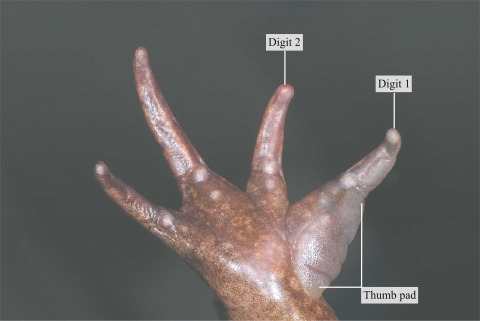
2) Place a frog on a dissection tray. To determine the frog's sex, look
at the hand digits, or fingers, on its forelegs. A male frog usually has
thick pads on its "thumbs," which is one external difference
between the sexes, as shown in the diagram below.


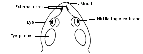
_ mouth
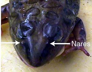
_ external nares
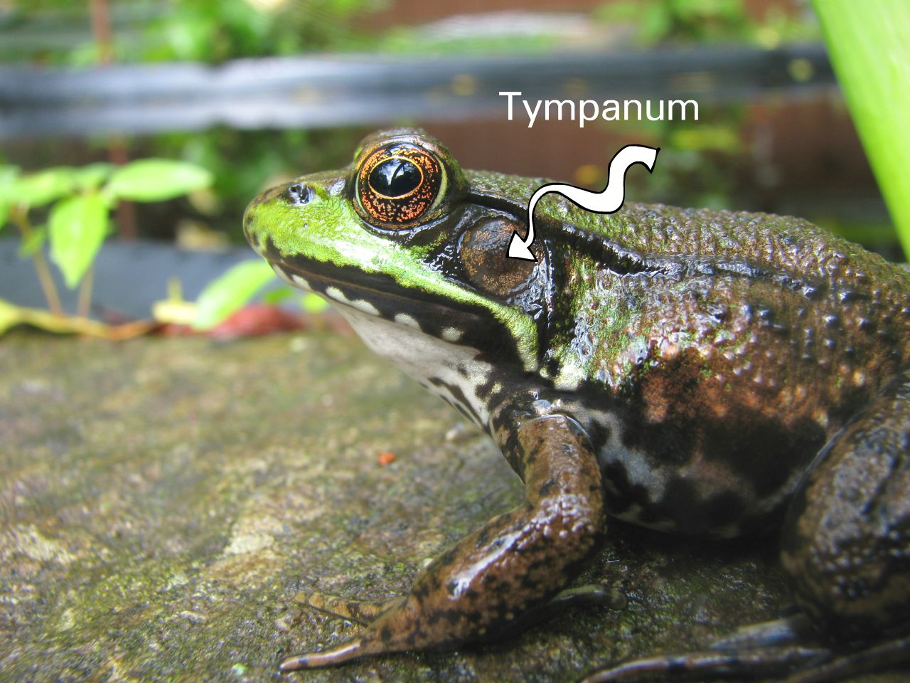
_ tympani
_ eyes

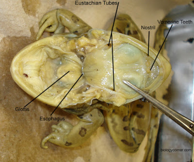
_ vomerine and maxillary teeth
_ internal nares
_ tongue
_ esophagus


Layer 1:
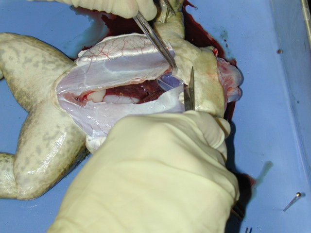
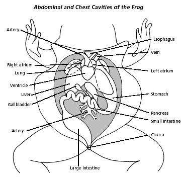
_ Liver - large, brownish organ covering most of the body cavity
_ Heart - small triangular organ between front legs and above liver


Layer 2: Lift liver with forceps to see
_ Gall Bladder - small greenish sac under liver
_ Stomach - large, firm sac on left side
_ Small Intestine - long, folded tube smaller than and above stomach


Layer 3
_ Lungs - small, reddish organs on either side of the heart
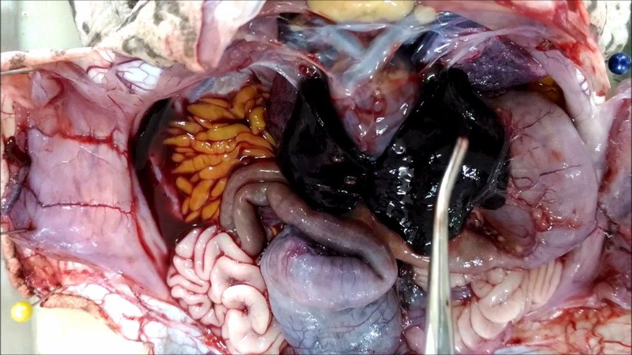
_ Pancreas - thin, yellowish ribbon between stomach and small intestine. Lift stomach to view.


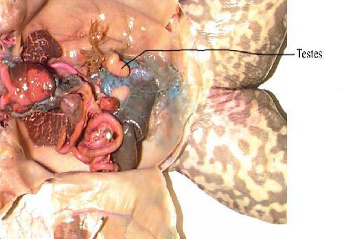

_ Testes - tan, bean shaped organs
_ Ovaries - dark organ which may fill most of the body cavity
11) Clean all equipment and your work area. Wash your hands.